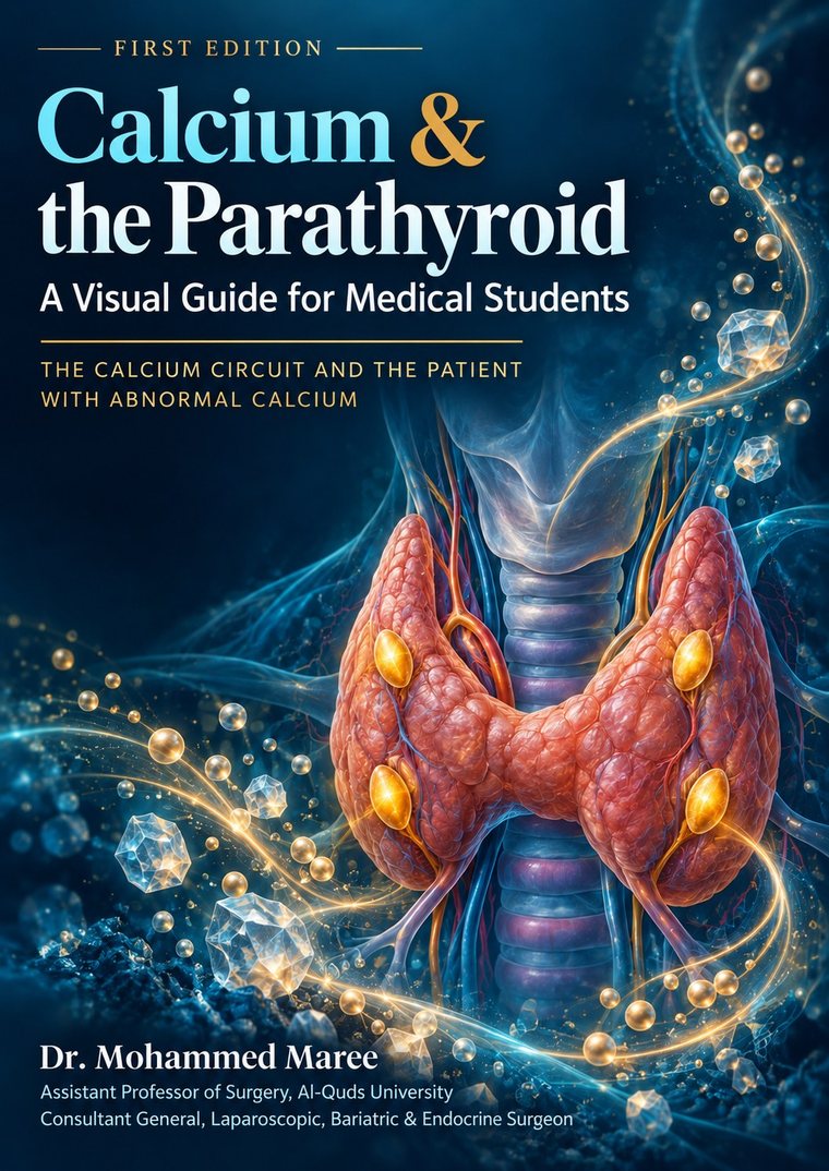
Calcium is one of the few things the body defends minute by minute. It is held inside a band roughly one and a half milligrams per decilitre wide, and it is defended by four glands that together weigh less than a paperclip.
Almost every calcium problem a surgeon meets — the woman with recurrent stones, the man whose bones ache, the patient whose fingers tingle the night after a thyroidectomy — is that defence system either working too hard or not working at all. Read the circuit once and the clinical half stops being a list to memorise: every decision in it is one step of the circuit failing.
DOI: 10.5281/zenodo.23158993 · Licensed CC BY-NC 4.0 · Volume 2 of Visual Guides for Medical Students
What is inside
Part 1 · The calcium circuit
- Where the calcium is
- The thermostat: the calcium-sensing receptor
- Four glands, two origins
- The signal: parathyroid hormone
- PTH on bone
- PTH on the kidney: calcium in, phosphate out
- PTH on the kidney: making the active vitamin D
- Calcitriol on the gut
- Closing the loop
- The whole circuit on one page
Part 2 · The patient
- Reading a calcium result
- Three numbers, one diagnosis
- The patient with hypercalcaemia: history
- The patient with hypercalcaemia: examination
- What the imaging shows
- Primary hyperparathyroidism: the surgical disease
- Localisation, and the operation
- Hypercalcaemia of malignancy, and the crisis
- Secondary and tertiary hyperparathyroidism
- The tingling fingers: hypocalcaemia after neck surgery
- Five questions at the bedside
Part 3 · Five patients
- The stone that led to the neck — a preoperative diagnosis, worked step by step
- The PTH that did not fall — in the operating theatre
- The operation that should never happen — familial hypocalciuric hypercalcaemia
- Day one after a total thyroidectomy — hypocalcaemia
- Eighteen months after the new kidney — tertiary hyperparathyroidism
The four milestones
- M1 — Describes the calcium circuit and names the actions of PTH
- M2 — Interprets a calcium, PTH and phosphate panel and reaches a diagnosis
- M3 — Assesses a patient with hypercalcaemia: history, examination, first-line investigations
- M4 — Acts as an intern: recognises hypercalcaemic crisis and post-thyroidectomy hypocalcaemia, starts first-line treatment, and escalates appropriately
A note on units
All values in the book are given in mg/dL, matching the chemistry report handed to you on the ward, with the conversion to mmol/L alongside. The reference intervals are printed in full on page 17 — check them against your own laboratory's sheet before quoting them in an exam or a clinic letter. The corrected calcium calculator on this site works in either set of units.
Who it is for
Medical students on a surgical or endocrine rotation, students preparing for an OSCE, interns, and junior surgical trainees. It assumes no prior knowledge and builds from the physiology upwards. Read Part 1 before the endocrine tutorial, and Part 2 alongside the thyroid and neck-lump station.
How to cite it
Maree M. Calcium & the Parathyroid: A Visual Guide for Medical Students. 1st ed. Visual Guides for Medical Students, Volume 2. Zenodo; 2026. doi:10.5281/zenodo.23158993
Read it online instead
Using it in teaching
The licence is CC BY-NC 4.0. You may share, print and adapt it for non-commercial teaching, provided the author is credited. If you use it with a student group, I would be glad to hear how it went.